拉曼光谱应用于生命科学
拉曼光谱已经成功应用于人体、动物和植物细胞与组织的分析。
在许多生命科学应用中都可以使用这种无标记分析技术获取化学和结构信息。
- 生物加工
- 蛋白质/肽结构分析
- 微生物学
- 体外和体内给药
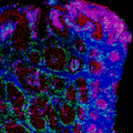
- 癌症研究/病理学
- 氧化还原生物学
- 再生医学
- 老龄化和神经退行性疾病
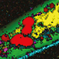
- 生物燃料和农业研究
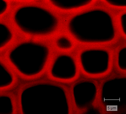
- 脂类组学
- 代谢组学
- 发育生物学
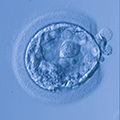
- 生殖生物学
- 病毒学
欢迎点击下方链接,了解我们如何在生命科学应用方面帮助您:
下载文档


拉曼光谱已经成功应用于人体、动物和植物细胞与组织的分析。
欢迎点击下方链接,了解我们如何在生命科学应用方面帮助您: