生命科学
拉曼光谱已经成功应用于人体、动物和植物细胞与组织的分析。
- 生物处理工艺
- 蛋白质/缩氨酸结构分析
- 微生物学
- 体外和体内给药
- 癌症研究/病理学
- 氧化还原生物学
- 再生医学
- 老龄化和神经退行性疾病
- 生物燃料和农业研究
- 脂类组学
- 代谢组学
- 发育生物学
- 生殖生物学
- 病毒学
欢迎点击下方链接,了解我们如何在生命科学应用方面帮助您:
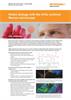
网络研讨会 — 用于研究氧化还原生物学的共振拉曼光谱
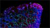
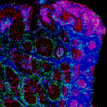
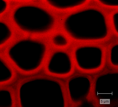
共振拉曼 (RR) 光谱是研究氧化还原生物学的理想工具。它不仅表现出对血红素蛋白的高灵敏度,还可以原位表征它们的氧化和氧合状态(溶液、细胞器、细胞和组织)。共振拉曼成像可提供化学和空间信息,将血红素蛋白分布、氧化状态和蛋白/细胞功能关联起来。
观看网络研讨会资料下载:生命科学
-
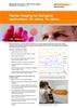
 样本: 利用拉曼光谱和成像技术进行生物分析
我们窥探微观世界的能力决定了生物研究领域的广度和深度。肉眼观察显微镜下的生物样本大有可为,但是利用拉曼光谱技术,我们可以超越视觉进入分子领域......甚至不止于此! 欢迎下载此样本,详细了解雷尼绍拉曼系统可大显身手的各种生物学应用。
-
 Application note: Redox biology with the inVia confocal Raman microscope [it]
Raman spectroscopy is sensitive to the presence of haem proteins and is ideal for studying their redox biology, without the need for isolation or staining. The redox of haem proteins is closely linked to their protein functions – oxygen transport and storage, electron transport, and scavenging of free radicals. By using Raman spectroscopy to elucidate redox states within biological systems, researchers can study redox dynamics and its effects on health regulation and diseases.
-
 Application note: Raman imaging for biological applications. No stains. No labels. [en]
Raman spectroscopy is an information-rich, label-free, non-invasive imaging technique that is ideal for life sciences research. It uses laser light scattering to provide a chemical fingerprint at each point of the analysed area and identifies the molecules present in samples.
-
 Product note: Microplate mapping with Renishaw Raman system's [en]
Renishaw’s microplate mapping package enables researchers to use Renishaw’s Raman spectroscopy products to rapidly and easily analyse material contained in microplates.
-
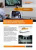
Raman chemical imaging for life sciences [en]
Short movie to demonstrate the benefits of using the inVia confocal Raman microscope for powerful, flexible, Raman Imaging for life sciences applications.