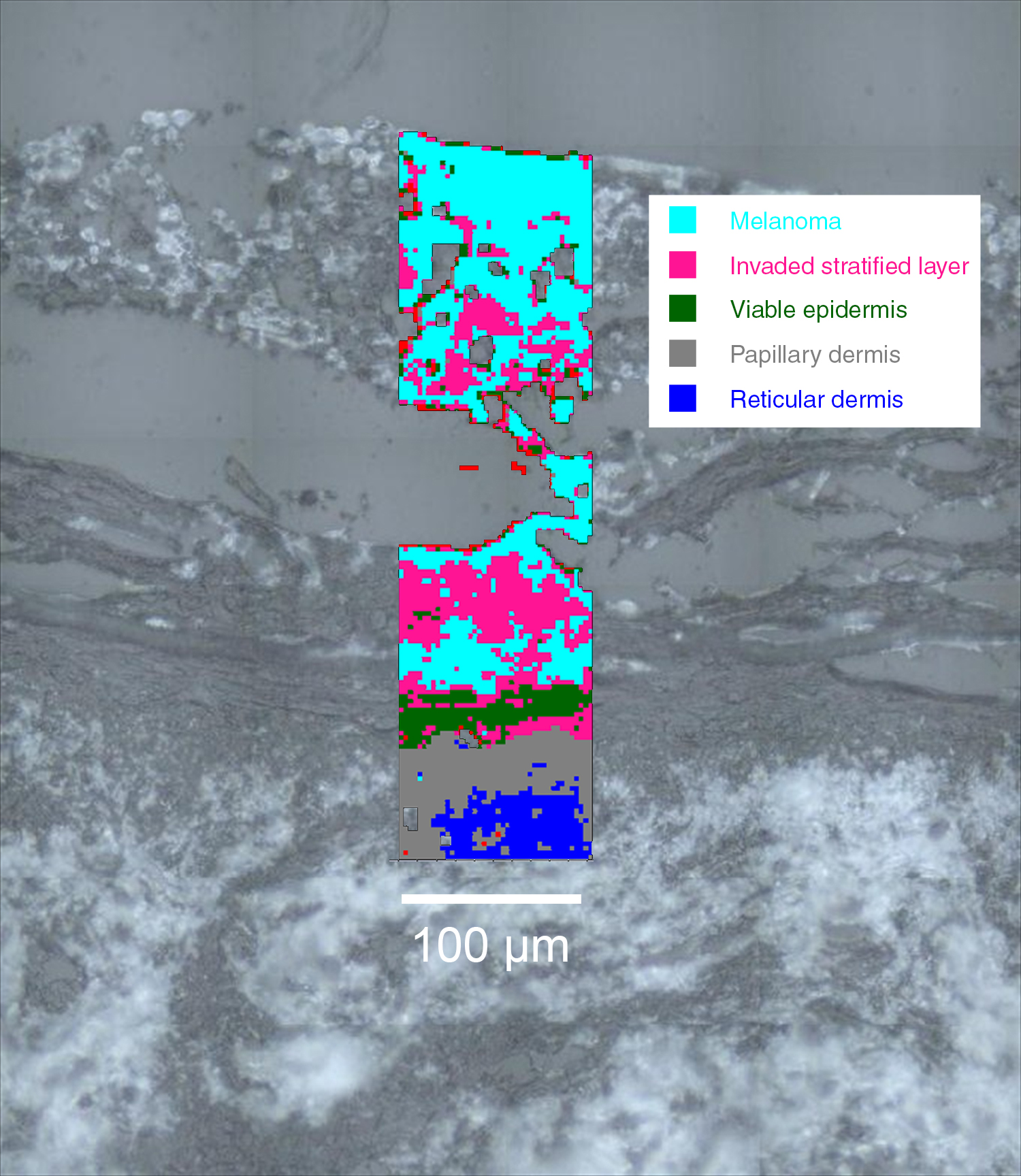
利用拉曼光谱分析生物组织
拉曼光谱作为一种无损、无标记的技术,是分析生物组织的理想工具。
您可以从核酸、蛋白质和脂类等实体中提取化学信息的完整光谱,而无需靶向生物分子、标记物、着色剂或染料。与蛋白质印迹法、气相色谱/质谱 (GC/MS)、基质辅助激光解吸电离飞行时间质谱 (MALDI-TOF) 等许多其他分析技术不同,拉曼分析不需要将样品均匀化。
快速、准确地识别组织层
区分、识别和界定癌前病变组织、癌变组织和健康组织。
- 无需与抗体结合:在优化方案时,可节省时间和金钱
- 基于组织的整体分子组成,可靠地界定并客观地识别组织中的解剖层次,并划定肿瘤边界。
- 避免主观的比色法和基于形态学的分析
- 识别形态上尚未发生改变的组织是否发生了化学变化(例如,DNA/RNA、糖原、脂类、蛋白质、脂类相的水平和DNA完整性)
了解生物系统
对组织进行完整的化学分析,并了解组织发生改变的潜在机制。
研究:
- 有机体的发育
- 疾病的发病机制
- 组织对药物或兴奋剂(例如,化疗药物、毒素和抗炎药)的反应
通过一次分析便可检测:
- 蛋白质、脂类、核酸和碳水化合物的水平和构象变化
- 矿物沉积的存在(例如,乳腺组织和动脉粥样硬化中的钙化)
- 血红蛋白的氧化还原态(例如,神经血红蛋白和肌红蛋白)
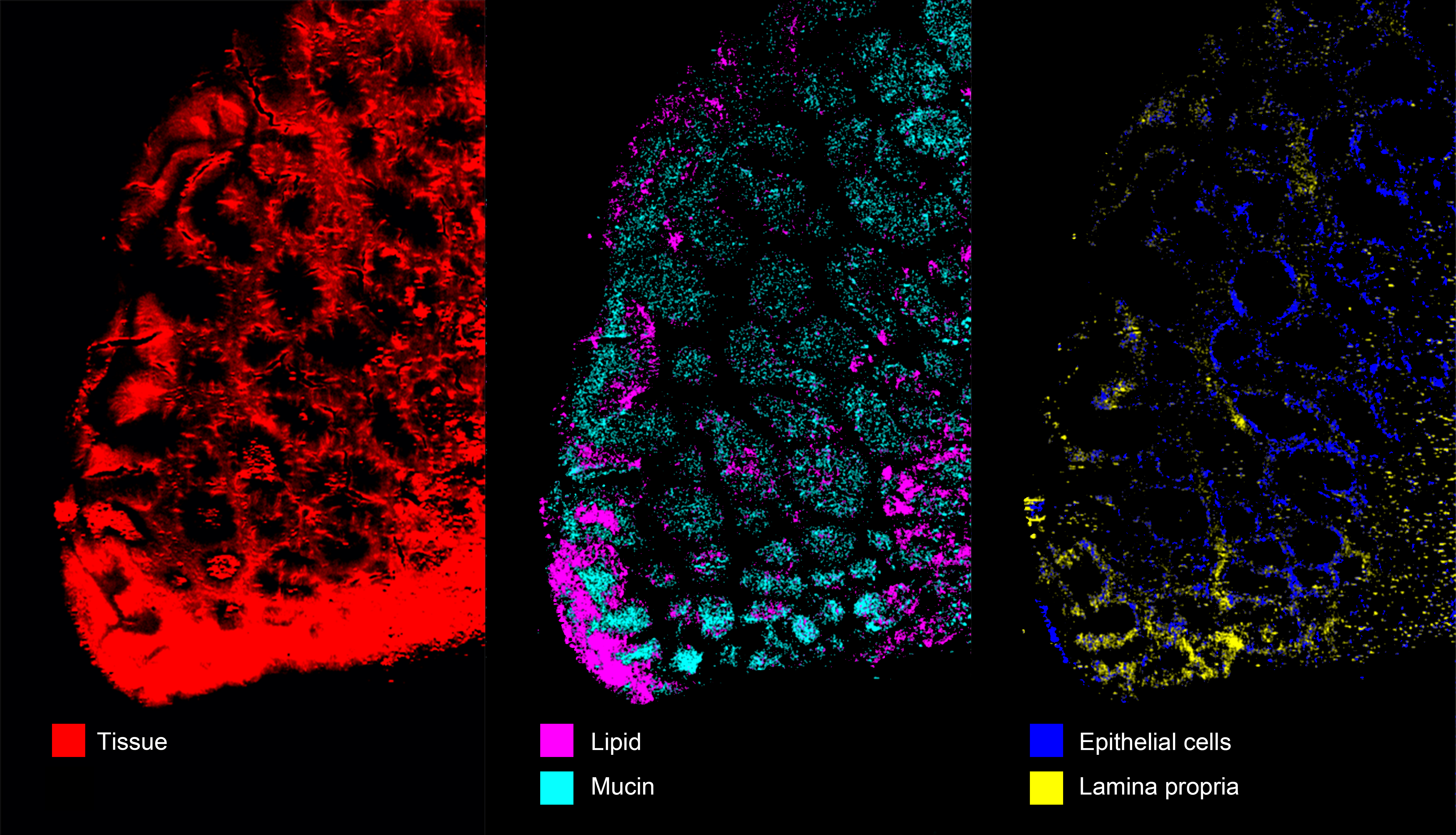
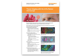
生成组织的图像
雷尼绍的StreamLine™(大面积快速成像)技术特别适合生成组织的图像。它采用线聚焦技术,允许使用高激光功率,同时可避免对组织造成光热损伤。这样可提高信号水平,并在尽可能短的时间内生成图像。
研究凹凸不平的表面
利用StreamLine的Surface成像模式,您可以生成样品的拉曼图像,即使是凹凸不平的样品也不例外。
自动化载玻片扫描
您可以将雷尼绍的全自动化拉曼系统配置为按顺序扫描多个组织切片,无需人工干预。这样不但节省了时间,而且能够提高拉曼系统的运行效率。
下载文档
网络研讨会 —利用拉曼光谱预测生物流体和组织中的疾病
拉曼光谱已成为医学领域的一项关键分析技术。它可以在不改变样品的原始状态的情况下,提取详细的化学信息。因此,拉曼光谱能够有效评估生物流体和组织内的疾病状态。
在本场网络研讨会中,我们将展示雷尼绍结构紧凑、易于使用的RA816生物分析仪如何将拉曼光谱应用于临床。它可以从体外样品中快速采集大量光谱,从而帮助您研发模型,用于预测和检测生物流体和组织中的疾病。
观看网络研讨会您或许对以下文献感兴趣:
Kast et al (2014) J Neurooncol doi 10.1007/s11060-014-1536-9
Bonifacio et al (2010) Analyst 135: 3193-3204
相关新闻报道
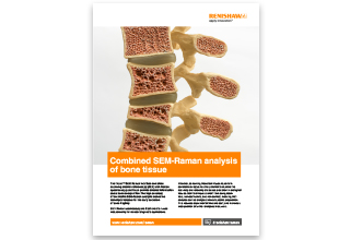
拉曼光谱具有识别骨关节炎患者病变软骨的潜力
由Riana Gaifulina博士领导的一组来自伦敦大学学院 (University College London)、英国皇家兽医学院 (Royal Veterinary College) 和查林十字医院 (Charing Cross Hospital) 的研究人员和临床医生已证明,拉曼光谱可以测定软骨磨损腐蚀的程度和骨关节炎疾病的整体状态。这项研究结果可能有助于骨关节炎退行性病变的及早发现与治疗。
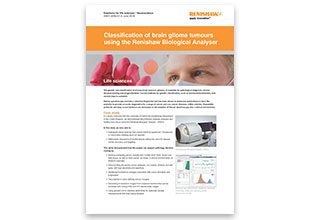
利用拉曼光谱分析生物组织,实现脑胶质瘤快速分类
研究人员使用雷尼绍RA816生物分析仪,在外科手术过程中鉴别出脑胶质瘤的不同基因亚型。
通过拉曼光谱检测在癌症治疗期间辐射对细胞和组织的损伤
一支跨学科的科学家和工程师团队正在使用拉曼光谱技术了解癌症治疗中使用的辐射对细胞和组织造成的损伤。
密歇根儿童医院利用拉曼光谱进行疾病研究
密歇根儿童医院 (Children’s Hospital of Michigan) 与韦恩州立大学 (Wayne State University) 共同利用雷尼绍inVia共焦显微拉曼光谱仪研究儿童期疾病。