联用拉曼系统
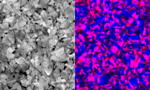
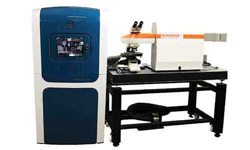
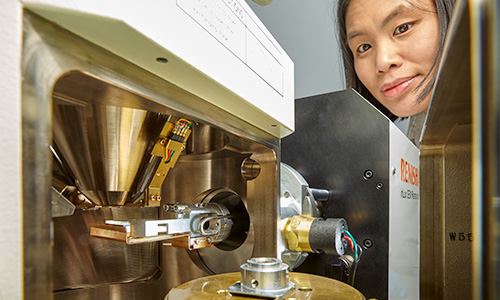
将雷尼绍的inVia™共焦显微拉曼光谱仪或Virsa™拉曼分析仪与其他分析仪器结合使用,可实现多模态成像能力。雷尼绍的拉曼光谱仪已成功与多种分析技术整合,包括配备针尖增强拉曼光谱 (TERS) 技术的原子力显微镜 (AFM)、扫描电子显微镜 (SEM)、荧光寿命显微成像 (FLIM)、纳米压痕技术,以及红外 (IR) 热成像。此外,雷尼绍拉曼仪器还可配置用于光致发光 (PL) 和光电流成像。
通过高效的工作流程,您可以在一台集成式仪器上利用两种或更多表征技术分析样品。使用雷尼绍的关联式显微光谱仪系统,您能够同时采用多种技术对同一点进行分析。下文详细介绍了我们的联用拉曼系统。
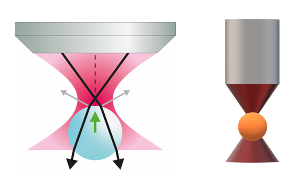
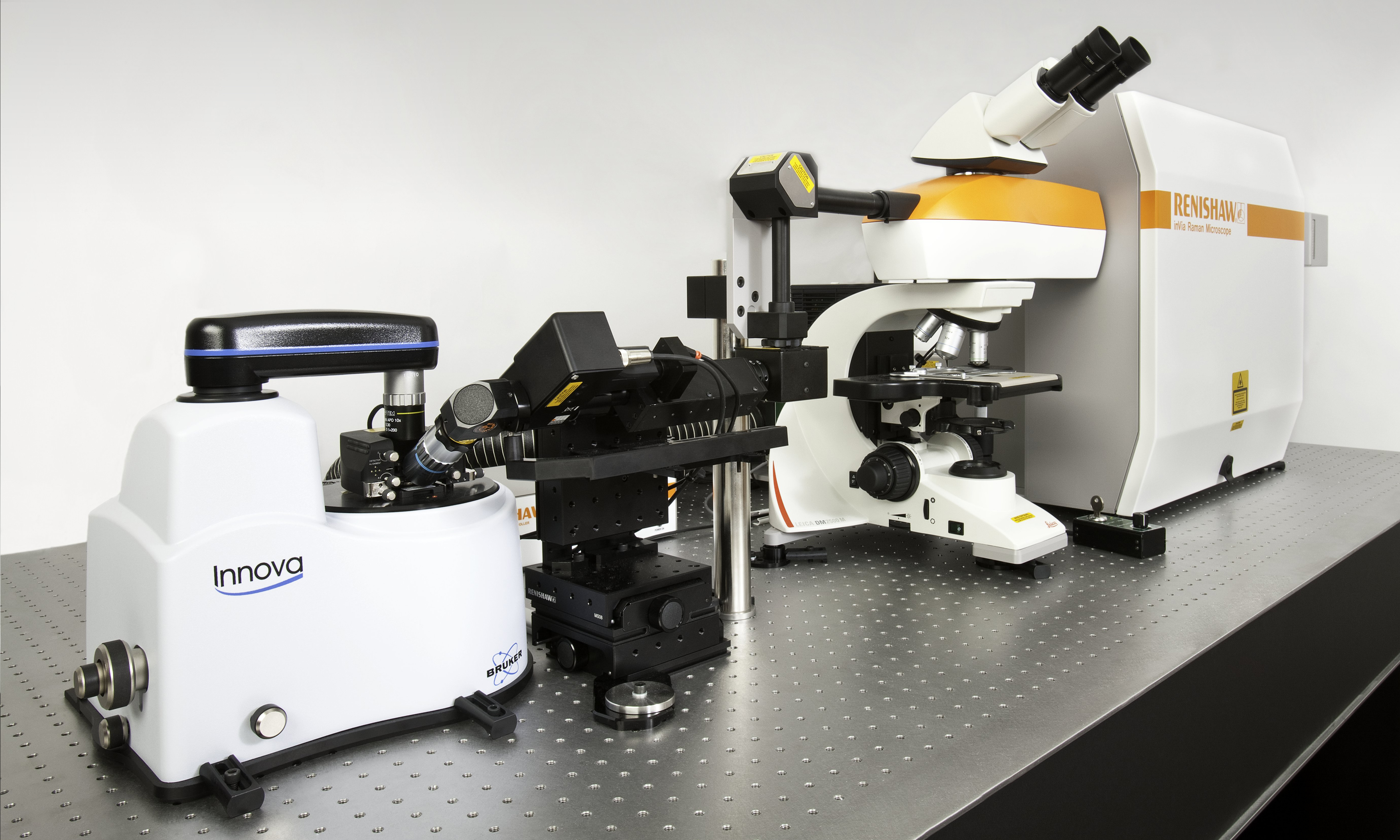
SPM/AFM-拉曼联用系统
您可以将inVia显微拉曼光谱仪与多种扫描探针显微镜 (SPM) 和原子力显微镜 (AFM) 结合使用,以揭示包括形貌和力学性能在内的互补性信息。通过添加针尖增强拉曼光谱 (TERS) 技术,还可实现纳米级化学分辨率。
了解更多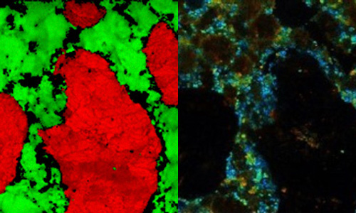
荧光寿命显微成像 (FLIM)
FLIM可与inVia显微拉曼光谱仪集成,以采集显示荧光基团的荧光寿命的空间图像。在细胞生物学中,FLIM可用于环境传感、分子相互作用监测,以及荧光基团识别等领域。
了解更多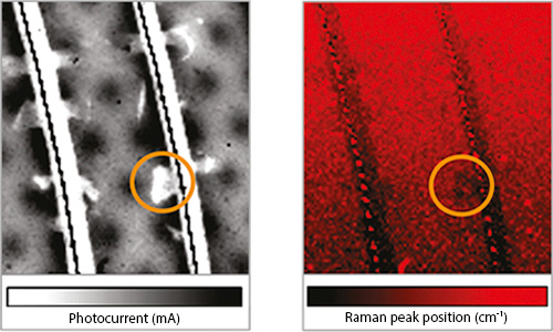
其他定制解决方案
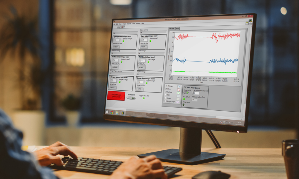
如果我们的标准产品不能完美贴合您的需求,我们的“非标准产品团队”能够以丰富的经验针对您的具体需求研发定制解决方案。详细了解拉曼光谱在同步辐射光束线和常规质量保证/质量控制 (QA/QC) 分析环境中的集成示例。
了解更多《光谱学》电子书:关联式拉曼成像技术的最新进展
拉曼光谱是一个迅速发展的领域,现代拉曼光谱仪为实验室提供了更高的易用性和灵敏度。了解如何将拉曼光谱仪与扫描电子显微镜 (SEM) 或荧光寿命成像显微镜 (FLIM) 结合使用,从而提升该技术并应用于不同领域。
- 拉曼仪器的发展和拉曼技术的创新;
- 在SEM样品室内进行原位拉曼光谱分析,以及inLux SEM-拉曼联用接口如何在SEM成像过程中提供互补性信息;
- 关联式FLIM和显微拉曼光谱技术,以及植物组织切片和HeLa细胞的成像应用示例。